Diagnostic and therapeutic strategies targeting the neurovascular-unit after organophosphate nerve agent exposure
Funded by Agence Nationale de la Recherche
Context and challenges
Organophosphate compounds, including pesticides and nerve agents, are highly toxic chemicals responsible for major global health and security concerns. They irreversibly inhibit cholinesterases, causing excessive acetylcholine accumulation that can lead to seizures, brain damage, blood brain barrier disruption, and neuroinflammation. Although current treatments exist, their efficacy in the central nervous system is limited, and the neurovascular consequences of exposure remain poorly understood, highlighting a critical gap in knowledge and therapy development.
Objective
The NeuroTarget project aims to characterize neurovascular alterations induced by nerve agents to identify new diagnostic biomarkers and therapeutic targets. We are using immuno-MRI to detect early vascular damage, and evaluating a novel immunotherapy targeting the tPA-NMDAR pathway to mitigate neurotoxicity, providing a proof of concept for improved diagnosis and treatment after nerve agent exposure.
Project coordinator
Cyrille Orset, PhD (U1237)
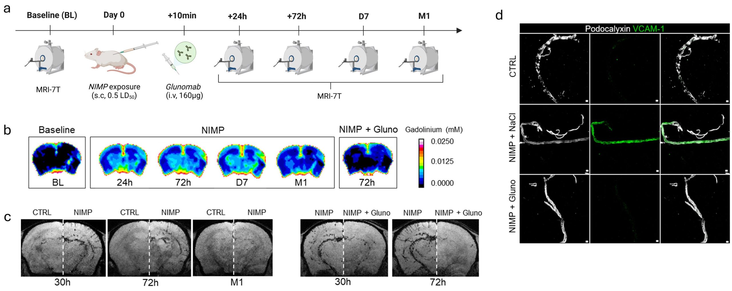
Blocking tPA-NMDAR as a therapeutic strategy following nerve agent (NA) exposure: Schematic representation of the experimental design (a). tPA–NMDAR signaling mediates blood-brain barrier (BBB) disruption induced by low-dose NA exposure, as shown by significant accumulation of gadolinium in the brain parenchyma at 24 hours, 72 hours, and even 7 days post-exposure (b). Since increased BBB permeability is closely linked to neurovascular inflammation, we evaluated endothelial activation using in vivo MRI with VCAM-1–targeted iron microparticles over a period of one month after NIMP exposure. In NIMP-intoxicated mice, neuroinflammation was observed in the somatosensory cortex (c). Glunomab® (patent INSERM U1237, PhIND) treatment reduced endothelial activation (c, d).
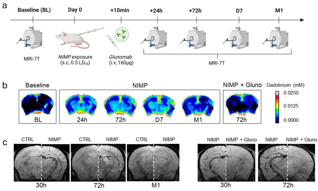
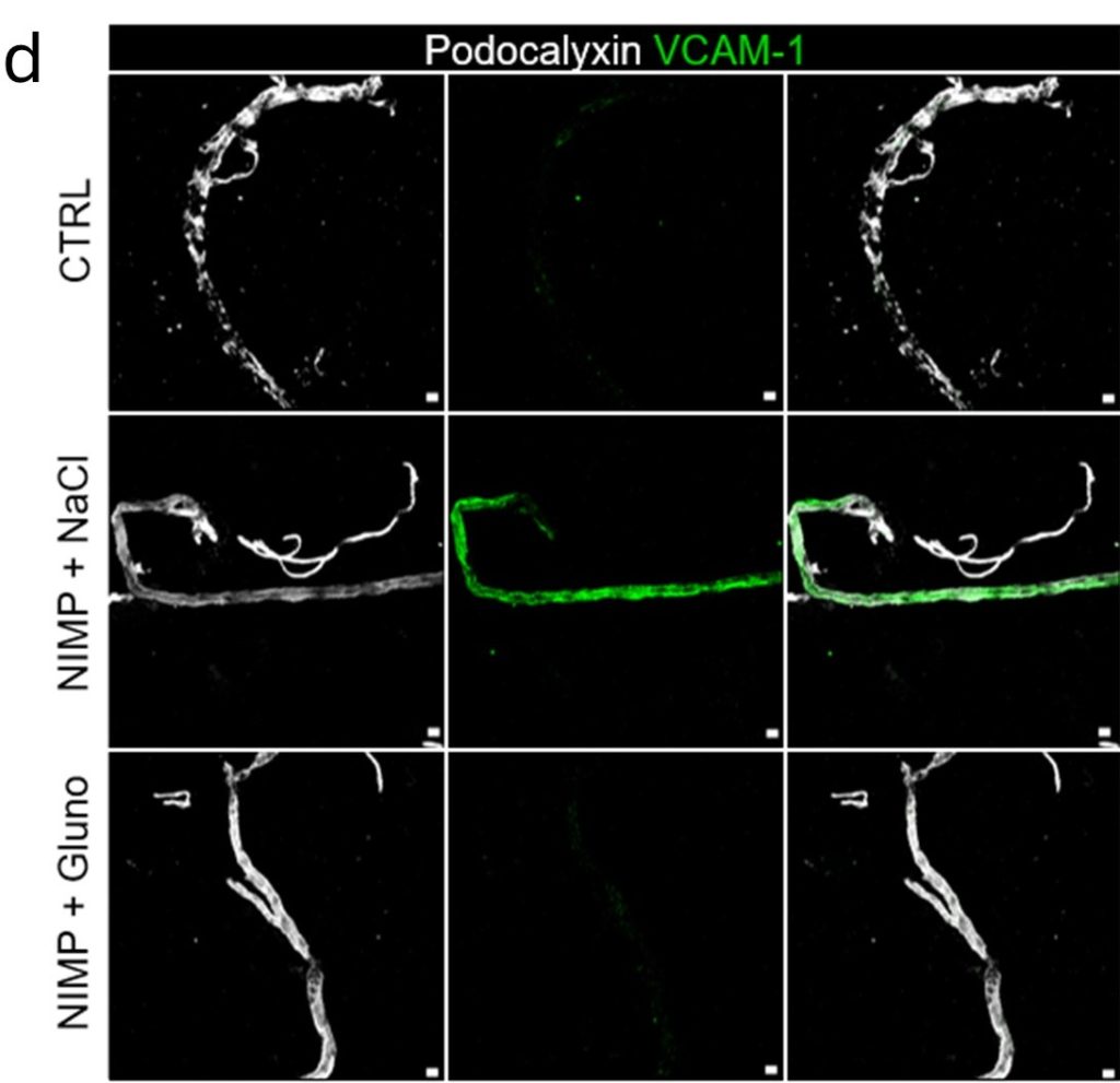
Blocking tPA-NMDAR as a therapeutic strategy following nerve agent (NA) exposure: Schematic representation of the experimental design (a). tPA–NMDAR signaling mediates blood-brain barrier (BBB) disruption induced by low-dose NA exposure (NIMP), as shown by significant accumulation of gadolinium in the brain parenchyma at 24 hours, 72 hours, and even 7 days post-exposure (b). Since increased BBB permeability is closely linked to neurovascular inflammation, we evaluated endothelial activation using in vivo MRI with VCAM-1–targeted iron microparticles over a period of one month after NIMP exposure. In NIMP-intoxicated mice, neuroinflammation was observed in the somatosensory cortex (c). Glunomab® (patent INSERM U1237, PhIND) treatment reduced endothelial activation (c, d).