Biomedical engineering for Neurovascular Disorders
INSERM U1237 – Physiopathology and imaging of neurological disorders (PhIND)
Brain Blood & Memory @ Caen-Normandie Institute (BBM@C)
📍Caen, Normandy, France



Our research expertise
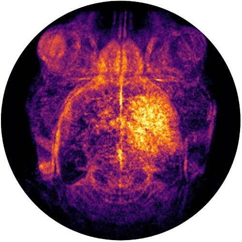
Starting in 2028, BiND aims to officially become a research team within PhIND-BBM@C, a structure led by Prof. Denis Vivien. Our group has strong expertise in biomaterials, vascular biology, animal models, radiopharmacy, and molecular imaging. In particular, we develop innovative immuno-imaging technologies (immuno-MRI, immuno-MPI, immuno PET/MRI, immuno-photoacoustics imaging) that enable non-invasive visualization, with exceptional sensitivity, of proteins involved in immune responses across different organs. To achieve this, we are continuously working on designing new probes capable of specifically targeting activated endothelial cells to detect leucocyte trafficking.

BiND co-directors
Maxime Gauberti, MD, PhD
Sara Martinez de Lizarrondo, PhD
What is immuno-imaging?
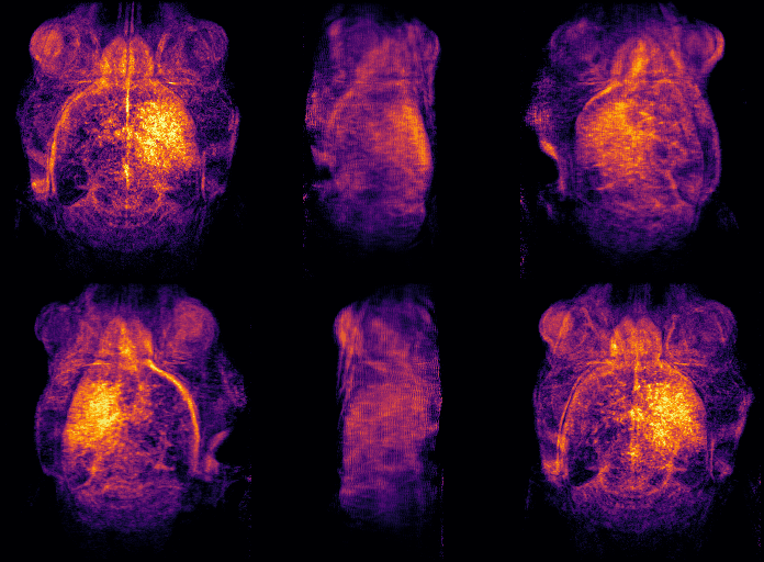
Under physiological conditions, the vascular endothelium is in a quiescent state characterized by a limited interaction with circulating leukocytes. Upon injury, the endothelial cells shift to an activated phenotype, involving increased adhesion molecules expression, such as P-Selectin, E-selectin, VCAM-1, ICAM-1, and MAdCAM-1, on their luminal surface. These proteins are involved in the initial recruitment of leukocytes and in their firm adhesion to the endothelial wall. By conjugating imaging contrast agents to specific antibodies, we can essentially “see” the expression of these adhesion molecules and reveal inflammatory processes. We synthesize biodegradable microsized ferromagnetic particles that enable the imaging of these processes with high sensitivity (Gauberti et al., 2018, Theranostics). We have named this approach “immuno-imaging“, to describe this powerful technology and its wide range of applications across different imaging modalities, which include early diagnosis, detecting subclinical injury, monitoring therapeutic response, and grading the severity of inflammatory disorder.


Non-invasive detection of inflammation using biodegradable particles
Acquisitions comparing a healthy control (sham) and a sepsis mouse model (induced by intraperitoneal injection of LPS) following injection of targeted contrast agent developed in our laboratory. In healthy animals, particles accumulate in the monocyte macrophague system (liver); in contrast, septic mice show a strong signal detected in the lungs and kidneys.
Meet the BiND team
We are a young and dynamic research group, passionate about developing cutting-edge imaging strategies to study and reveal the hidden dynamics of inflammation in various disease contexts. We are driven by the need to create solutions that address clinical needs and can be translated to patients.
📍Caen, Normandy, France

State-of-the-art in vivo imaging and research platforms
PhIND benefits from a unique combination of laboratories and equipment with access to cutting-edge imaging technologies through the Cyceron platform, including:
- Imaging facility: 7T MRI, hybrid PET-MRI scanner, magnetic particle imaging (MPI), photoacoustic imaging, functional ultrasound imaging (fUS).
- Molecular and cellular biology labs.
- Microscopy facilities : wide-field and confocal microscopy, calcium imaging, spinning disk, and STED microscopy.
- Radiochemistry and radiopharmaceutical labs.
- Central animal facility.
- Surgical suite: setup of various preclinically relevant models .