






Project coordinator: Sara Martinez de Lizarrondo, PhD
Cardiovascular disease is the leading cause of death worldwide. Commonly used imaging methods, such as echocardiography and cardiac MRI, are essential for diagnosis and monitoring. However, these conventional techniques only show the consequences of cardiac dysfunction, without providing molecular insights into the underlying pathological processes. This project (finished in 2026) aimed to assess the potential of immuno-MRI for cardiac imaging, enabling the visualization of heart–immune cell interactions and uncovering the inflammatory processes that drive major cardiac pathologies.
This project has received funding from the Normandy Region under the “Projets Emergents” program and is co-funded by the European Union through the European Regional Development Fund (FEDER).
Thanks to this funding, we have successfully established cardiac molecular MRI in our laboratory, enabling in vivo molecular imaging of the heart. “MAGENTA” allowed us to support a postdoctoral position in our lab, acquire the equipment necessary for microparticle production, and successfully evaluate the performance of this contrast agent in preclinical models of common cardiac pathologies, including myocardial infarction, myocarditis, and anthracycline-induced cardiotoxicity (manuscript under preparation).
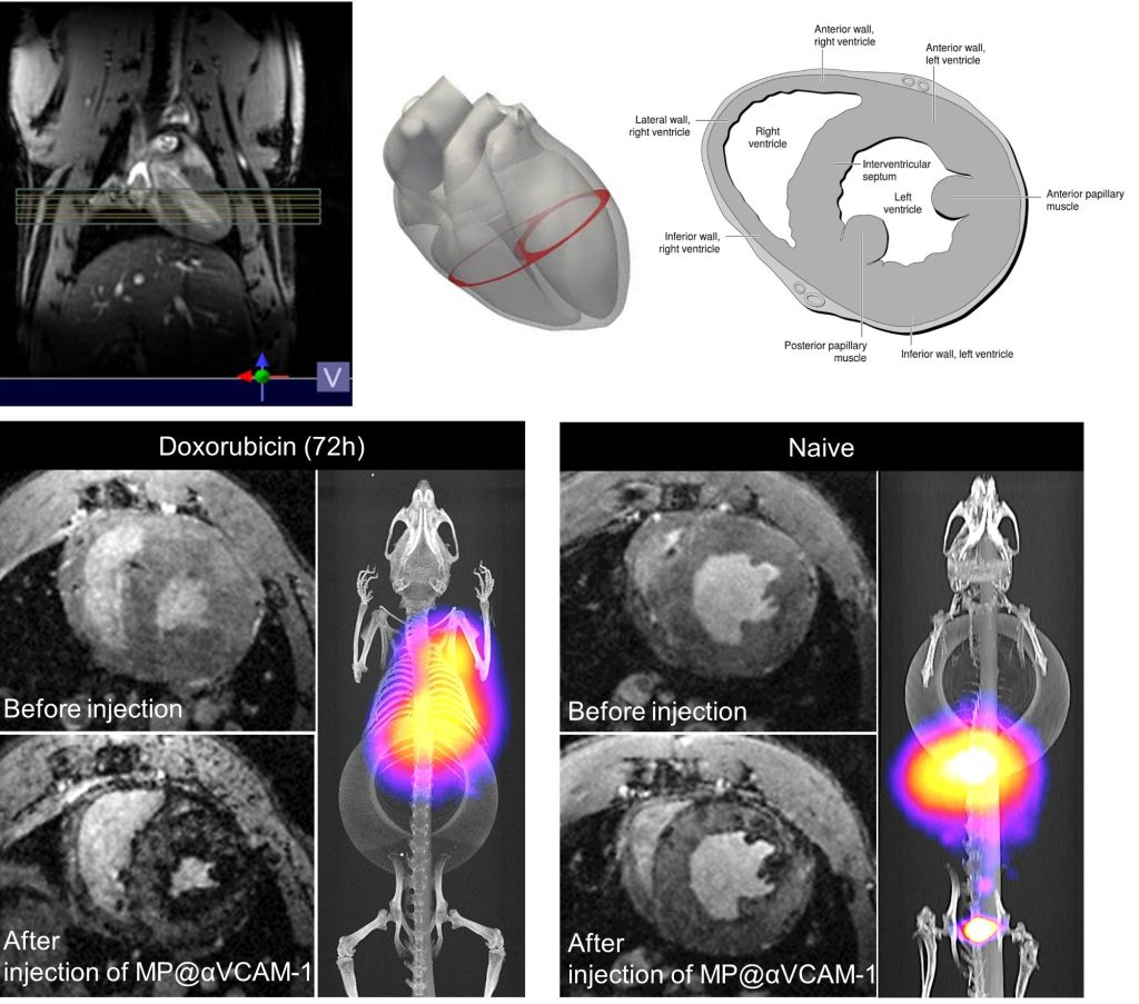
First line: short-axis views of the heart, the slice reveal the right and left ventricles as well as the left ventricular myocardium and cavity.
Second line: Myocardial inflammation induced by doxorubicin, a commonly used chemotherapeutic agent in clinic. Anthracycline-induced cardiotoxicity affects a significant proportion of patients and can lead to left ventricular dysfunction and heart failure. With increasing long-term cancer survival, there is growing interest in early detection of these late-onset cardiac complications. In this study, a mouse model received a single intraperitoneal injection of doxorubicin. Immuno-MRI and immuno-MPI using microparticles targeting VCAM-1 (MP@VCAM-1) as contrast agent revealed a clear inflammatory signal in the myocardium, whereas in control (naïve) animals the signal was restricted to the liver, the physiological clearance site of the contrast agent.
Project coordinator: Abraham Martín, PhD (Achucarro Basque Center for Neuroscience Fundazioa, Spain)
Local project partner: Maxime Gauberti, PhD
Because of the ageing population, the prevalence of stroke is expected to dramatically increase in coming years: by 2025, 1.5 million European people will suffer a stroke each year. The therapeutic window of thrombolysis and mechanical thrombectomy is generally limited, reducing the number of patients who can benefit from them. For this reason, there is an urgent need for effective treatments to improve the clinical management of subacute ischemic stroke. During secondary ischemic injury, neurons die and inflammatory signals attract immune cells into the brain. This process is worsened by damage to the blood–brain barrier (BBB), which becomes more permeable due to harmful enzymes (among them, metalloproteases) released by activated immune cells such as microglia and neutrophils. This project (finished in 2026), aimed to develop theragnostic tools aiming at measuring and modulating metalloproteases MMPs activity, monitoring immune cell trafficking across the BBB and measuring BBB permeability during the subacute phase of stroke.
This project has received funding from the Network of European Funding for Neuroscience Research
Thanks to this support, we have strengthened collaboration between the participating centers (Caen, Achucarro, Budapest, and Berlin), and we are currently organizing the first European multicenter preclinical trial in immuno-MRI for the unbiased validation of our biodegradable probes for ultrasensitive imaging.