Diagnosis of pulmonary embolism in pregnant women using magnetic particle imaging
Funded by Fondation Recherche Cardio-Vasculaire
Programme de recherche « DANIELE HERMANN – COEURS DE FEMMES »
Context and challenges
Pulmonary embolism is the third most common cardiovascular disease and remains a major cause of maternal morbidity and mortality. Current diagnostic imaging techniques, including computed tomography pulmonary angiography and ventilation/perfusion scintigraphy, rely on ionizing radiation, raising important safety concerns for women, particularly during pregnancy, where both maternal breast tissue and the fetus may be exposed.
Objective
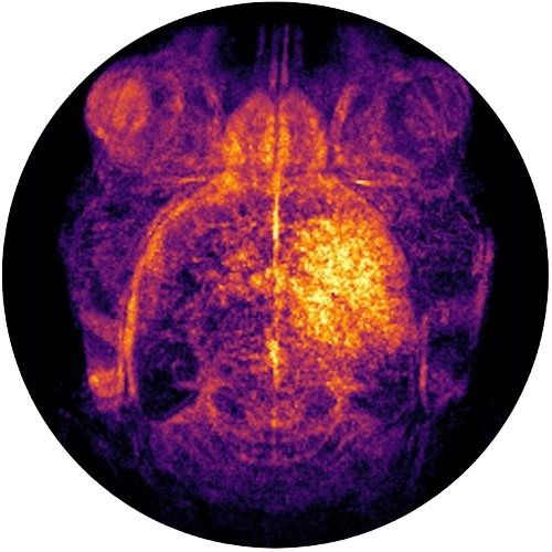
This project aims to develop a novel, radiation-free imaging approach for the diagnosis of pulmonary embolism in women using Magnetic Particle Imaging. Inspired by conventional lung perfusion scintigraphy, the strategy consists of engineering superparamagnetic iron oxide particle aggregates that can transiently lodge within the pulmonary microvasculature and be detected by MPI. this work seeks to establish a safer and highly sensitive imaging modality for the diagnosis and monitoring of pulmonary embolism, particularly in pregnant women.
Project coordinator
Sara Martinez de Lizarrondo, PhD (U1237)
Related links
Fondation Recherche Cardio Vasculaire
Sara Martinez de Lizarrondo – Lauréate 2024 bourse « cœurs de femmes »

Our lab is home to the only MPI device in France and one of the first in the world
Our lab is equipped with all the necessary facilities to carry out this project, from particle synthesis to in vivo imaging of experimental models of pulmonary embolism. The team is recognized in the field of imaging and the production of biocompatible superparamagnetic contrast agents.
Acknowledgment to Fondation Recherche Cardio Vasculaire
We gratefully acknowledge the support of the Fondation Recherche Cardio-Vasculaire through its Cœurs de Femmes research program, which provided funding for this project
